DYKDDDDK tag
FUJIFILM Wako provides antibodies, antibody coupling beads, and peptides to purify and detect DYKDDDDK tag fusion proteins.
Application Data
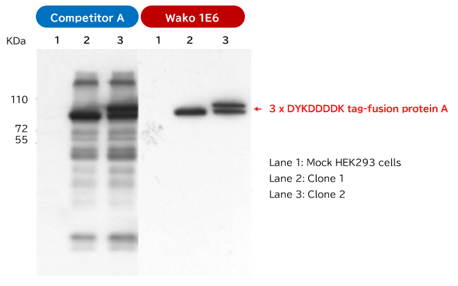
Comparison of specificity by Western blotting

Dr. Shuichi Kusano, Research Field: Division of Persistent & Oncogenic Viruses, Center for Chronic Viral Diseases, Graduate School of Medicine and Dentistry, Kagoshima University.
A plasmid expressing 3 x DYKDDDDK-tagged fusion protein A was transfected into HEK293 cell lines. Cloned cells were randomly selected after incubation for 24 hours.
Selected cell clones were lysed with RIPA Buffer. The 3 x DYKDDDDK-tagged fusion protein was detected using Western blotting on the cell lysate obtained.
As primary antibodies, FUJIFILM Wako's antibody (clone No. 1E6, Product Number: 018-22381) and competitor's antibody were used in Western blotting.
Compared to Competitor A, our antibody (clone No. 1E6) can specifically recognize the DYKDDDDK sequence.
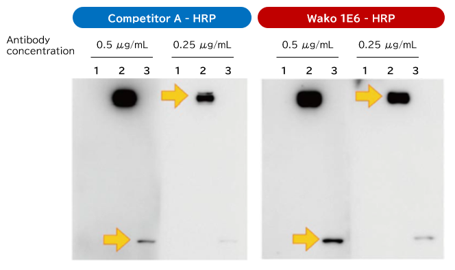
Comparison of sensitivity by Western blotting

Lane 1: 293T cell lysate
Lane 2: 293T cell lysate transiently expressing a DYKDDDDK tagged protein A
Lane 3: 293T cell lysate transiently expressing a DYKDDDDK tagged protein B
Blocking: TBS-T that contains 5% skim milk, overnight at 4 °C
Antibody reaction: 2 hr at room temperature
Luminescence reagent: Immunostar®LD
Detection: LAS-3000, exposure time 15 minutes
1E6-HRP shows more sensitivity than Competitor A
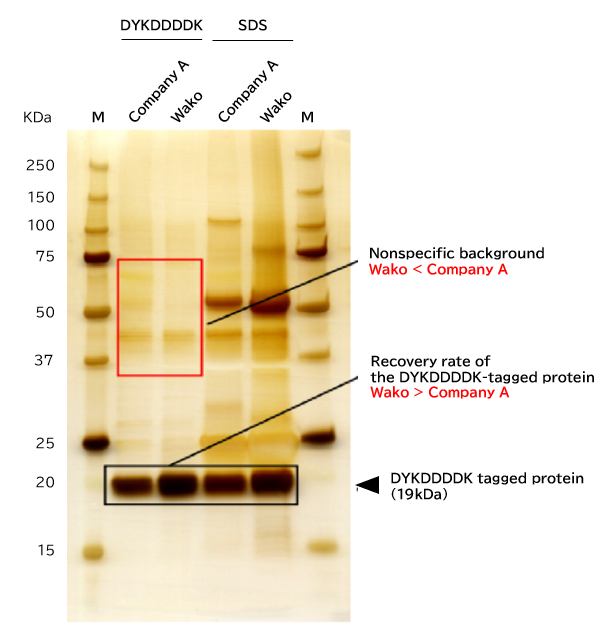
Antigen recovery performance of Anti-DYKDDDDK tag Antibody coupled Agarose Beads

In comparison to Competitive product A, this antibody performed as well, or better, in terms of antigen recovery.
Elution methods
DYKDDDDK: Competitive elution with DYKDDDDK peptide
SDS: Denaturing elution with 2% SDS
The amount of beads used
Anti-DYKDDDDK tag Antibody Beads (Wako): 20 µL/assay
Affinity beads (Company A): 20 µL/assay
The amount of antigen added
E. coli cell lysate containing the DYKDDDDK tagged fusion protein: 20 mg/assay
Immunoprecipitation conditions
3 hours at 4° C
Elution methods
150 µg/mL DYKDDDDK peptide (Product Number: 047-34581): 20 µL/assay, followed by incubation for 30 minutes at 4℃
2% SDS sample buffer: 20 µL/assay, followed by boiling for 5 minutes.
SDS-PAGE
Sample volume: 10 µL
Detection
Silver stain
An E.coli lysate, over-expressing the DYKDDDDK-tagged fusion protein (approximately 19kDa), was prepared. After immunoprecipitation using this product and Competitive product A, the tagged protein was eluted with the DYKDDDDY peptide and SDS. The obtained target protein was separated by electrophoresis and an antigen recovery efficiency was compared by silver staining.
Reactivity of Anti DYKDDDDK tag Antibody Magnetic Beads

Regardless of tag location, allows for the recovery of recombinant proteins
After adding each DYKDDDDK-tagged fusion protein to cell lysates, immunoprecipitation was performed. The antigen recovery was compared using Western blotting.
After performing SDS-PAGE with a 4% SDS sample buffer, luminescence intensity was detected with peroxidase-labeled antibody (Product Number: 015-22391) by Western blotting.
M:Marker Protein
Met:N-terminal Met-DYKDDDDK-BAP
N:N-terminal DYKDDDDK-BAP
C:C-terminal DYKDDDDK-BAP
Product List
- Open All
- Close All
Anti DYKDDDDK tag, Monoclonal Antibodies
Labeled Anti DYKDDDDK tag, Monoclonal Antibodies
Anti DYKDDDDK tag, Monoclonal Antibody (Agarose Beads)
Anti DYKDDDDK tag, Monoclonal Antibody (Magnetic Beads)
DYKDDDDK Peptide
DYKDDDDK-BAP Peptides
3×DYKDDDDK Peptide
For research use or further manufacturing use only. Not for use in diagnostic procedures.
Product content may differ from the actual image due to minor specification changes etc.
If the revision of product standards and packaging standards has been made, there is a case where the actual product specifications and images are different.
The prices are list prices in Japan.Please contact your local distributor for your retail price in your region.


