Fluorescent-labeled rBC2LCN (Animal-derived-free)
This product is a fluorescently labeled recombinant lectin, rBC2LCN (AiLecS1), which shows strong affinity for undifferentiated cells.
rBC2LCN is produced in E. coli and corresponds to the N-terminal domain of BC2L-C, a lectin derived from Burkholderia cenocepacia. BC2LCN binds with exceptionally high affinity to H-type 3 (Fucα1-2Galβ1-3GalNAc), a mucin-type O-glycan present on the surface of undifferentiated human ES/iPS cells. Accordingly, it serves as a useful marker for identifying undifferentiated human ES/iPS cells.
Because this product is fluorescently labeled, undifferentiated human ES/iPS cells can be detected in live cultures simply by adding it to the medium at an appropriate concentration. It is compatible with both cell staining and flow cytometry. Furthermore, no cytotoxicity has been observed, even after several days of culture in the presence of this product.
This product is free of animal-derived components. Therefore, it is suitable for confirming undifferentiated status or removing undifferentiated human ES/iPS cells by flow cytometry during the manufacture of regenerative medicine products derived from human ES/iPS cells.
Two fluorescent labels are available: Green (FITC) and Red (Cy5 region).
Features
- Animal-derived-free : manufactured without animal-derived raw materials
- Enables cell staining simply by adding to the culture medium
- Allows clear staining of live cells without fixation
- Suitable for both cell staining and flow cytometry
- Low cytotoxicity, allowing continued culture after staining
- Recognizes cell-surface glycans, similar to Tra-1-60 and SSEA-4
Product Information
- Sterility tested
- Supplied in PBS(-) solution
- Recommended dilution
Live-cell staining: 1:100 – 1:1,000
Flow cytometry: 1:100 – 1:1,000 - Excitation / Emission wavelengths:
| Excitation | Emission | |
|---|---|---|
| rBC2LCN-FITC | 495 nm | 520 nm |
| rBC2LCN-635 | 634 nm | 654 nm |
Instructions for Use
Staining Live Cells (Live-cell Imaging)
- Prepare human ES/iPS cells in culture.
- Add 1–10 μL of rBC2LCN-FITC, AF or rBC2LCN-635, AF per 1 mL of culture medium.
- 37 ℃, 5 % CO2for 30 minutes.
- Replace the medium with fresh medium, HBSS(+), or D-PBS(-).
- Observe using a fluorescence microscope.
Staining Fixed Cells
- Prepare human ES/iPS cells in culture.
- Remove the culture medium and wash cells with HBSS(+).
- Add 4 % paraformaldehyde (PFA) solution and incubate at room temperature for 10–20 minutes.
- Remove PFA and wash three times with D-PBS(-).
- Add D-PBS(-).
- Add 1–10 μL of rBC2LCN-FITC, AF or rBC2LCN-635, AF per 1 mL of D-PBS(-) .
- Incubate at 37 ℃, 5 % CO₂ for 30 minutes.
- Replace with D-PBS(-).
- Observe using a fluorescence microscope.
Flow Cytometry
- Prepare human ES/iPS cells in culture.
- Dissociate cells into single-cell suspension using a cell dissociation reagent.
- Transfer the cell suspension to a tube and centrifuge at 1,000 rpm for 3 minutes.
- Discard the supernatant, resuspend cells in FCM Buffer*, centrifuge again, and discard the supernatant.
*FCM Buffer: D-PBS(-) or HBSS(-), optionally supplemented with 10 mmol/L EDTA and 1 % BSA or FBS. - Resuspend cells in FCM Buffer at 5x10⁶ cells/mL.
- Add 1–10 μL of fluorescently labeled rBC2LCN per 1 mL of cell suspension.
- Incubate for 30 minutes at room temperature in the dark.
- Centrifuge at 1,000 rpm for 3 minutes and discard the supernatant.
- Resuspend cells in FCM Buffer, centrifuge again, and discard the supernatant.
- Resuspend cells in an appropriate volume of FCM Buffer.
- Analyze by flow cytometry.
【Precautions for Use】
- Staining with rBC2LCN can be maintained for 2–3 days.
- When using serum-containing media, background fluorescence may increase.
- When sorting cells by flow cytometry, add Y-27632 during cell dissociation and to the FCM buffer at a final concentration of 10 μmol/L.
Staining Live Human iPS Cells (Live-Cell Imaging)
Human iPS cell line 201B7 was stained without fixation using rBC2LCN, SSEA-4, and Tra-1-60. Images were acquired 2 hours after staining. Compared with staining using the other antibodies, rBC2LCN enabled clearer visualization of the cells.
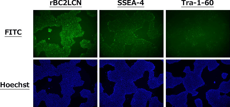
(Dilution ratio: 1:100)
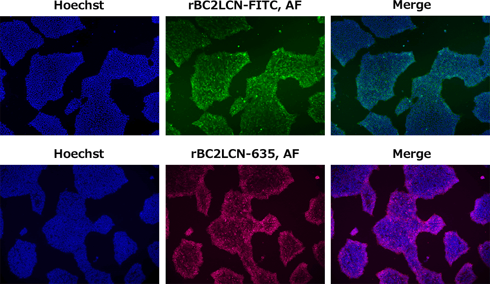
Fixed-Cell Staining of Human iPS Cells
Human iPS cell line 201B7 was fixed with paraformaldehyde and stained with rBC2LCN, Oct3/4, and DAPI. Compared with live-cell staining, the cells were stained more clearly.
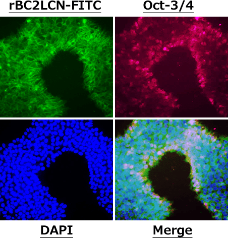
(Dilution ratio: 1:100)
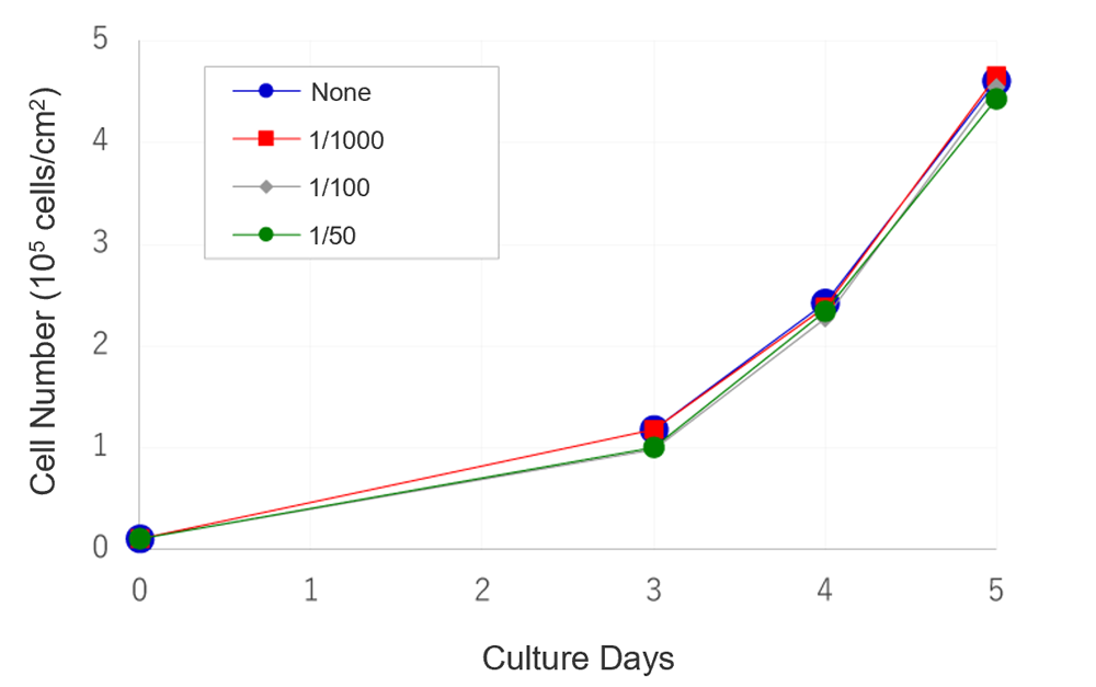
Cytotoxicity Evaluation in Human iPS Cells
Human iPS cell line 201B7 was cultured with rBC2LCN-FITC, AF at 1/1,000, 1/100, and 1/50 of the culture medium volume. Cell proliferation was comparable to the untreated control at all concentrations of rBC2LCN-FITC, AF. In addition, cell morphology remained similar between treated and untreated cells.
-
-
〔Cell line〕
Human iPS cell line 201B7〔Culture medium〕
StemSure® hPSC Medium Δ + 35 ng/mL bFGF〔Coating〕
Matrigel® hESC-Qualified Matrix〔Seeding density〕
4×10⁴ cells/well (12-well plate)
Isolation of Human iPS Cells by Flow Cytometry
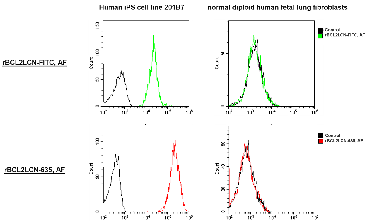
Human iPS cell line 201B7 and normal diploid human fetal lung fibroblasts were stained with rBC2LCN-FITC, AF or rBC2LCN-635, AF and analyzed by flow cytometry. The results show that undifferentiated human iPS cells can be distinguished from differentiated human diploid fibroblasts.
References
- Watanabe, T., Yasuda, S., Kusakawa, S., Kuroda, T., Futamura, M., Ogawa, M., Mochizuki, H., Kikkawa, E., Furukawa, H., Nagaoka, M. and Sato, Y.: Cytotherapy, 23, 176 (2021). Multisite studies for validation and improvement of a highly efficient culture assay for detection of undifferentiated human pluripotent stem cells intermingled in cell therapy products.
- Haramoto, Y., Onuma, Y., Mawaribuchi, S., Nakajima, Y., Aiki, Y., Higuchi, K., Shimizu, M., Tateno, H. and Hirabayashi, J.: Regen. Ther., 14, 306 (2020). A technique for removing tumourigenic pluripotent stem cells using rBC2LCN lectin.
- Saito, A., Hiemori, K., Kiyoi, K. and Tateno, H.: Sci. Rep., 8, (2018). Glycome analysis of extracellular vesicles derived from human induced pluripotent stem cells using lectin microarray.
- Tateno, H. and Saito, S.: Molecules, 22 (2017). Engineering of a Potent Recombinant Lectin-Toxin Fusion Protein to Eliminate Human Pluripotent Stem Cells.
- Tateno, H., Hiemori, K., Hirayasu, K., Sougawa, N., Fukuda, M., Warashina, M., Amano, M., Funakoshi, T., Sadamura, Y., Miyagawa, S., Saito, A., Sawa, Y., Shofuda, T., Sumida, M., Kanemura, Y., Nakamura, M., Okano, H., Onuma, Y., Ito, Y., Asashima, M. and Hirabayashi, J.: Regen. Ther., 6, 1 (2017). Development of a practical sandwich assay to detect human pluripotent stem cells using cell culture media.
- Tateno, H., Onuma, Y., Ito, Y., Minoshima, F., Saito, S., Shimizu, M., Aiki, Y., Asashima, M ando Hirabayashi, J.: Stem Cell Reports, 4, 811 (2015). Elimination of Tumorigenic Human Pluripotent Stem Cells by a Recombinant Lectin-Toxin Fusion Protein.
- Tateno, H., Onuma, Y., Ito, Y., Hiemori, K., Aiki, Y., Shimizu, M., Higuchi, K., Fukuda, M., Warashina, M., Honda, S., Asashima, M. and Hirabayashi, J.: Sci. Rep. , 4, 4069 (2014). A medium hyperglycosylated podocalyxin enables noninvasive and quantitative detection of tumorigenic human pluripotent stem cells.
- Tateno, H., Matsushima, A., Hiemori, K., Onuma, Y., Ito, Y., Hasehira, K., Nishimura, K., Ohtaka, M., Takayasu, S., Nakanishi, M., Ikehara, Y., Ohnuma, K., Chan, T., Toyoda, M., Akutsu, H., Umezawa, A., Asashima, M. and Hirabayashi, J.: Stem Cells Transl. Med., 2, 265 (2013). Podocalyxin is a glycoprotein ligand of the human pluripotent stem cell-specific probe rBC2LCN.
- Onuma, Y., Tateno, H., Hirabayashi, J., Ito, Y. and Asashima, M.: Biochem. Biophys. Res. Commun., 431, 524 (2013). rBC2LCN, a new probe for live cell imaging of human pluripotent stem cells.
Product List
- Open All
- Close All
For research use or further manufacturing use only. Not for use in diagnostic procedures.
Product content may differ from the actual image due to minor specification changes etc.
If the revision of product standards and packaging standards has been made, there is a case where the actual product specifications and images are different.
The prices are list prices in Japan.Please contact your local distributor for your retail price in your region.
Please contact us via the inquiry form.