Anti P2RY12 Antibodies
P2RY12 is a G protein-coupled receptor (GPCR) whose primary ligand is ADP. In the central nervous system, it is specifically expressed in microglia, making it a widely used microglial marker in neuroscience research. Because its expression in macrophages is minimal, P2RY12 also serves as a valuable marker for distinguishing microglia from macrophages found around blood vessels in the brain. "Anti P2RY12, Guinea Pig" is a polyclonal antibody against P2RY12 derived from guinea pigs and it can be used in immunohistochemistry for co-staining with other marker antibodies, such as anti-Iba1.
What is P2RY12?
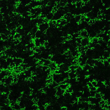
Anti-P2RY12, Guinea Pig (Product Code: 011-28873).
P2RY12 is a G protein-coupled receptor (GPCR) whose primary ligand is ADP. Coupled with Gi proteins, it inhibits adenylate cyclase upon ADP binding. Purinergic receptors are classified into P1 and P2 receptors: P1 receptors bind adenosine, while P2 receptors are further divided into P2X and P2Y receptors. P2X receptors are ligand-gated ion channels, whereas P2Y receptors are GPCRs. Among the P2Y receptor subtypes, P2RY12 is one example. It is also known by other names, such as P2Y12, P2Y12, or P2Y12R.
P2RY12 is specifically expressed by microglia in the central nervous system and is widely used as a microglial marker in neuroscience research. Previous studies suggest that microglia may detect neuronal damage in the brain through P2RY12-mediated ADP signaling. In peripheral tissues, P2RY12 is expressed by platelets, contributing to platelet aggregation and blood coagulation.
As mentioned earlier, P2RY12 is expressed by microglia but shows minimal expression in macrophages1). Therefore, co-staining with an anti-Iba1 antibody, which recognizes both microglia and macrophages, together with an anti-P2RY12 antibody, a microglia-specific marker, is effective for distinguishing microglia from macrophages found around blood vessels in the brain. Notably, P2RY12 is expressed in homeostatic microglia (previously referred to as "resting microglia"), but its expression is decreased in disease-associated microglia (DAM) and in microglia from Alzheimer’s disease model mice2-3).
Anti P2RY12, Guinea Pig
The “Anti P2RY12, Guinea Pig” is a guinea pig polyclonal antibody, raised against P2RY12. It can be used to perform multiplex immunohistochemistry.
Antibody Information
| Clonality | Polyclonal |
|---|---|
| Antigen | Synthetic peptide (P2RY12 C-terminal sequence) |
| Host | Guinea pig |
| Formulation | PBS, 0.05% Sodium azide |
| Conjugate | Unconjugated |
| Cross-reactivity | Mouse* |
| Application | Immunohistochemistry (frozen section) 1:500-2,000 |
Performance Data
Comparison with anti-Iba1 antibody
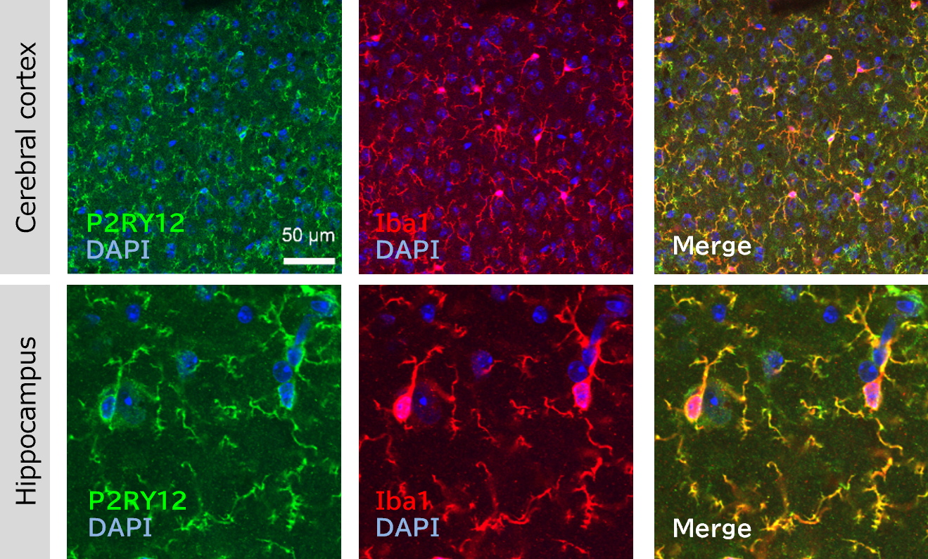
Immunohistochemistry was performed on mouse brain (frozen sections) using anti-P2RY12 and anti-Iba1 antibodies.
Data by courtesy of
Dr. Miyata, Department of Applied Biology, Kyoto Institute of Technology
Antibodies
P2RY12
Primary Antibody: Anti P2RY12, Guinea Pig (Product Number: 011-28873) 1:1,000
Secondary Antibody: Alexa Fluor® 488 AffiniPure Goat Anti-Guinea Pig IgG (H+L) (Jackson ImmunoResearch, Product Number: 106-545-003)
Iba1
Primary Antibody: Anti Iba1, Rabbit (for Immunocytochemistry) (Product Number: 019-19741) 1:1,000
Secondary Antibody: Alexa Fluor® 594-AffiniPure Goat Anti-Rabbit IgG (H+L) (Jackson ImmunoResearch, Product Number: 111-585-003)
[Result]
The co-localization of P2RY12 and Iba1 signals was confirmed.
Differentiating between microglia and macrophages
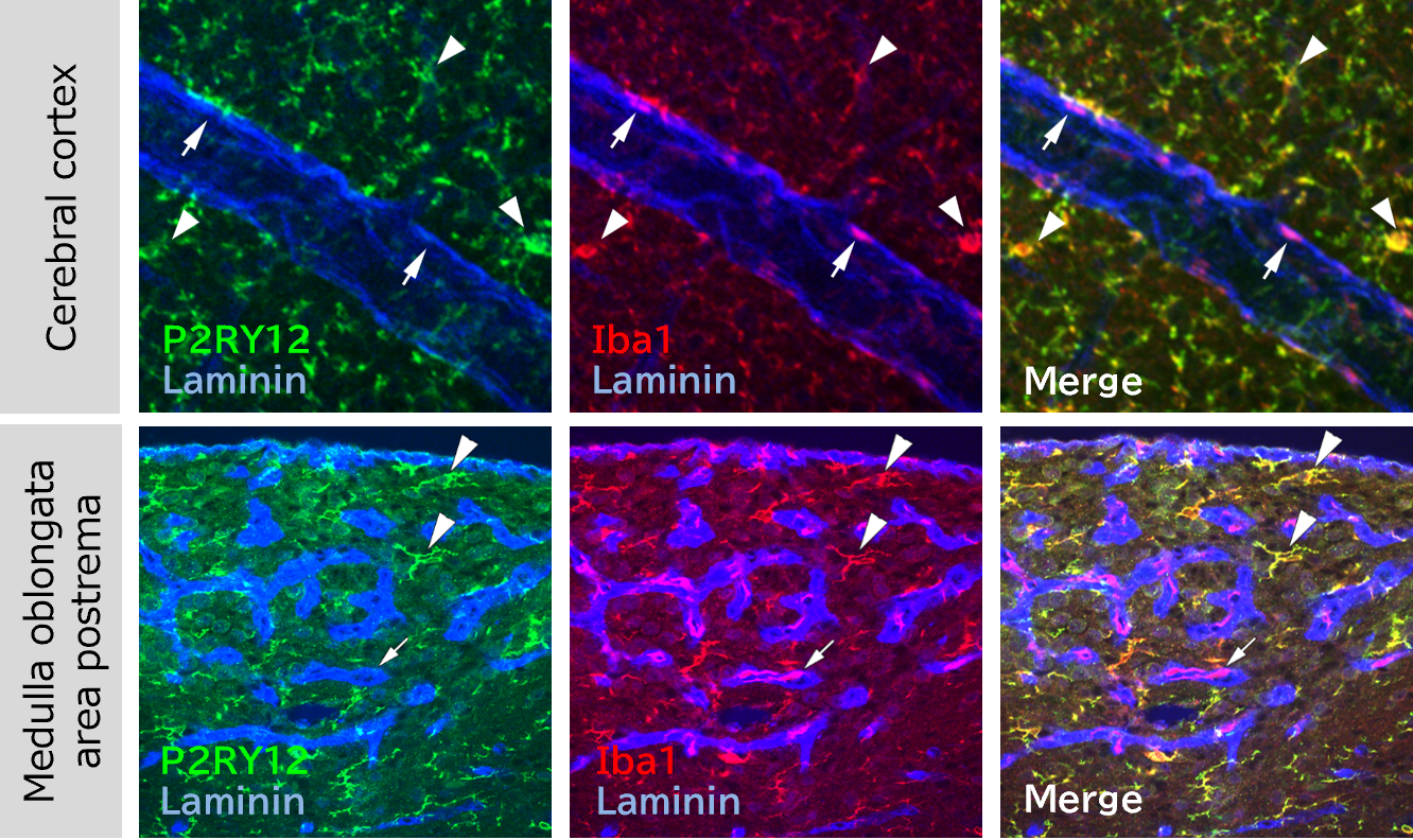
In the cerebral cortex and the medulla oblongata area postrema, triple staining was performed using anti-P2RY12 antibody (microglia marker), anti-Iba1 antibody (microglia/macrophage marker), and anti-laminin antibody (basement membrane marker of cerebral blood vessels), and the localization of cells was confirmed.
Data by courtesy of
Dr. Miyata, Department of Applied Biology, Kyoto Institute of Technology
Antibodies
P2RY12
Primary Antibody: Anti P2RY12, Guinea Pig (Product Number: 011-28873) 1:900
Secodary Antibody: Alexa Fluor® 488 AffiniPure Goat Anti-Guinea Pig IgG (H+L) (Jackson ImmunoResearch, Product Number: 106-545-003)
Iba1
Primary Antibody: Anti Iba1, Rabbit (for Immunocytochemistry) (Product Number: 019-19741) 1:500
Secondary Antibody: Alexa Fluor® 594-AffiniPure Goat Anti-Rabbit IgG (H+L) (Jackson ImmunoResearch, Product Number: 111-585-003)
Laminin
Primary Antibody: Anti Laminin Antibody (Rat, Made in Dr. Miyata’s Lab) 1:200
Secondary Antibody: DyLight™ 405-conjugated AffiniPure™ Goat Anti-Rat IgG (H+L) (Jackson ImmunoResearch, Product Number: 112-475-003)
[Result]
Among the Iba1-positive cells, those that are present in the brain parenchyma are P2RY12-positive and are thought to be microglia (arrowheads). On the other hand, Iba1-positive/P2RY12-negative cells are present around the blood vessel basement membrane, and it is thought that these cells are macrophages (arrows).
Comparison with anti-Iba1 antibody and anti-TMEM119 antibody
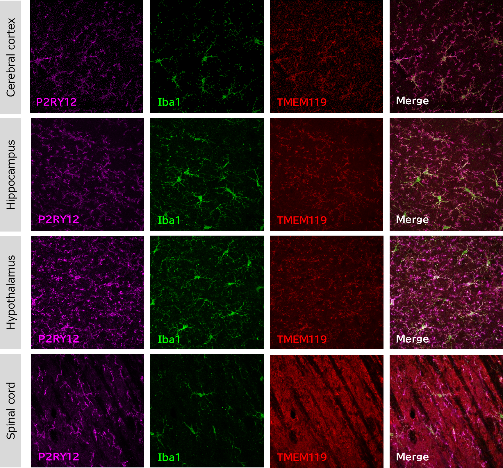
Immunohistochemistry was performed on mouse brain and spinal cord (frozen sections) using anti-P2RY12, anti-Iba1 and anti-TMEM119 antibodies
Antibodies
P2RY12
Primary Antibody: Anti P2RY12, Guinea Pig (Product Number: 011-28873) 1:1,000
Secondary Antibody: Alexa Fluor® 647 AffiniPure Donkey Anti-Guinea Pig IgG (H+L) (Jackson ImmunoResearch, Product Number: 706-605-148)
Iba1
Primary Antibody: Anti Iba1, Goat (Product Number: 011-27991) 1:1,000
Secondary Antibody: Alexa Fluor® 488 AffiniPure Donkey Anti-Goat IgG (H+L) (Jackson ImmunoResearch, Product Number: 705-545-147)
TMEM119
Primary Antibody: Anti-TMEM119 Antibody (Rabbit, conventional product) 1:100
Secondary Antibody: Alexa Fluor® 594-AffiniPure Donkey Anti-Rabbit IgG (H+L) (Jackson ImmunoResearch, Product Number: 711-585-152)
[Result]
Compared to anti-TMEM119 antibody, our anti-P2RY12 antibody staining was able to detect fine protrusion structures more clearly.
Comparison with conventional product
Immunohistochemical staining was performed on mouse brain and spinal cord (frozen sections) using our company's and conventional anti-P2RY12 antibodies.
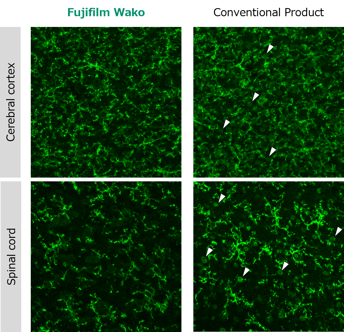
Antibodies
Fujifilm Wako
Primary Antibody: Anti P2RY12, Guinea Pig (Product Number: 011-28873) 1:1,000 (Antibody concentration 0.5 μg/mL)
Secondary Antibody: Alexa Fluor® 488 AffiniPure Donkey Anti-Guinea Pig IgG (H+L) (Jackson ImmunoResearch, Product Number: 706-545-148)
Conventional product
Primary Antibody: Anti-P2RY12 Antibod (Rabbit, conventional product) 1:200 (Antibody concentration 0.5 μg/mL)
Secondary Antibody: Alexa Fluor® 488 AffiniPure Donkey Anti-Rabbit IgG (H+L) (Jackson ImmunoResearch, Product Number: 711-545-152)
[Result]
With the conventional product, nonspecific signals thought to be from neurons were observed (arrowheads). On the other hand, with this product, almost no nonspecific signals were observed.
Additional application data and user feedback are available on the product details page.
References
- Butovsky, O. et al.: Neurosci., 17(1), 131(2014).
Identification of a unique TGF-β–dependent molecular and functional signature in microglia - Keren-Shaul, H. et al.: Cell, 169(7), 1276(2017).
A unique microglia type associated with restricting development of Alzheimer’s disease - Maeda, J. et al.: Brain Commun., 3(1), fcab011(2021).
Distinct microglial response against Alzheimer's amyloid and tau pathologies characterized by P2Y12 receptor
Product List
- Open All
- Close All
Anti P2RY12, Guinea Pig (polyclonal antibody)
For research use or further manufacturing use only. Not for use in diagnostic procedures.
Product content may differ from the actual image due to minor specification changes etc.
If the revision of product standards and packaging standards has been made, there is a case where the actual product specifications and images are different.
The prices are list prices in Japan.Please contact your local distributor for your retail price in your region.


