Anti Synapsin Antibodies
Synapsin is a synaptic vesicle-associated protein which is thought to regulate synaptic vesicle dynamics. Due to their predominant localization at nerve terminals, synapsins are widely used as presynaptic markers in immunohistochemistry.
Fujifilm Wako offers guinea pig anti-Synapsin I antibodies that are useful for multiplex staining.
What is Synapsin?
Synapsin is a synaptic vesicle-associated protein. In mammals, three synapsin genes have been identified: synapsin I, II, and III. Synapsin I and II are associated with synaptic vesicles containing neurotransmitters such as glutamate, GABA, dopamine, and acetylcholine, but are absent from vesicles containing neuropeptides. Synapsins are thought to regulate synaptic vesicle dynamics, with their function modulated through phosphorylation and dephosphorylation. Kinases such as CaMKI and CaMKII phosphorylate synapsins, whereas phosphatases such as calcineurin mediate their dephosphorylation.
Due to their predominant localization at nerve terminals, synapsins are widely used as presynaptic markers in immunohistochemistry.
Anti Synapsin I, Guinea Pig
The “Anti Synapsin I, Guinea Pig” is a guinea pig polyclonal antibody, raised against Synapsin I. It can be used to perform multiplex immunohistochemistry1-2).
Antibody Information
| Clonality | Polyclonal |
|---|---|
| Antigen | Rat Synapsin I |
| Host | Guinea pig |
| Formulation | Antiserum |
| Conjugate | Unconjugated |
| Cross-reactivity | Mouse, Rat |
| Application | Immunocytochemistry 1:1,000 Immunohistochemistry (Frozen Section) 1:1,500 |
Application Data
Immunohistochemistry
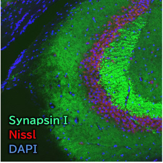
Species: Mouse
Site: Hippocampus (CA3)
Sample: Frozen section
Antibody concentration: 1:1,500
Data by courtesy of
Dr. Miyata, Department of Applied Biology, Kyoto Institute of Technology

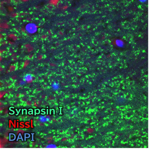
Species: Mouse
Site: Cerebral cortex (Layer V)
Sample: Frozen section
Antibody concentration: 1:1,500
Data by courtesy of
Dr. Miyata, Department of Applied Biology, Kyoto Institute of Technology
Immunocytochemistry
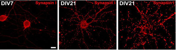
Sample: Rat hippocampus primary cultured neurons
Antibody concentration: 1:1,000
Data by courtesy of
Dr. Miyata, Department of Applied Biology, Kyoto Institute of Technology
[Result]
On day 7 of culture, only a few synapses were observed, whereas by day 21, numerous well-defined punctate synapses were evident. The cultured neurons are metabolically active, and the weak fluorescence observed in the cell bodies likely corresponds to synapsin I either being synthesized in the endoplasmic reticulum or transported along axons.
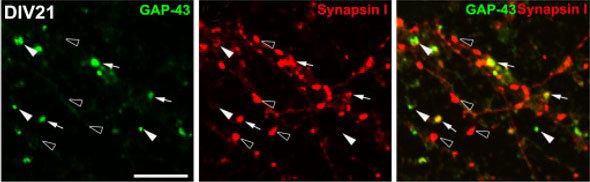
Sample: Rat hippocampus primary cultured neurons
Antibody concentration: 1:1,000
- Note:
- white arrows head GAP43 positive synapse
black arrows head Synapsin I positive synapse
Data by courtesy of
Dr. Miyata, Department of Applied Biology, Kyoto Institute of Technology
[Result]
Colocalization of GAP-43 and Synapsin I was observed at synapses (white arrows) in primary rat hippocampal neurons.
References
- Morita, S. and Miyata, S.: Cell Biochem. Funct., 31(5), 400(2013).
Synaptic localization of growth‐associated protein 43 in cultured hippocampal neurons during synaptogenesis - Kawai, S. et al.: Cell Biochem. Funct., 38(4), 392(2020).
Transient increase of microglial C1q expression in the circumventricular organs of adult mouse during LPS‐induced inflammation
Product List
- Open All
- Close All
Anti Synapsin I, Guinea Pig (Polyclonal)
For research use or further manufacturing use only. Not for use in diagnostic procedures.
Product content may differ from the actual image due to minor specification changes etc.
If the revision of product standards and packaging standards has been made, there is a case where the actual product specifications and images are different.
The prices are list prices in Japan.Please contact your local distributor for your retail price in your region.