Anti HuC/D Antibodies
Hu proteins are RNA-binding proteins which are thought to regulate mRNA stability and translational efficiency. Among the Hu proteins, HuB, HuC, and HuD are specifically expressed in neurons and contribute to neuronal differentiation and maintenance. In immunohistochemistry, anti-HuC/D antibodies are commonly used as neuron-specific markers.
Fujifilm Wako offers guinea pig anti-HuC/D antibodies that are useful for multiplex staining.
What is Hu proteins?
Hu proteins are RNA-binding proteins, and HuD was identified as the target antigen of autoantibodies present in the serum of patients who developed Paraneoplastic encephalomyelitis1). In vertebrates, four types -HuR, HuB, HuC, and HuD- have been identified, all of which contain three RNA-binding domains. Their structures are highly conserved, and the Drosophila ELAV (Embryonic Lethal Abnormal Vision) protein is a well-known homolog of the Hu proteins.
Hu proteins are thought to bind primarily to AU-rich elements in mRNA, thereby regulating mRNA stability and translational efficiency. Potential targets of Hu proteins include genes involved in cell proliferation, such as p21 and c-fos, as well as genes associated with neurite outgrowth, such as GAP-43 and Tau2).
Among the Hu proteins, HuB, HuC, and HuD are specifically expressed in neurons and contribute to neuronal differentiation and maintenance by regulating mRNA expression. In immunohistochemistry, anti-HuC/D antibodies are commonly used as neuron-specific markers.
Anti HuC/D, Guinea Pig
The “Anti HuC/D, Guinea Pig” is a guinea pig polyclonal antibody, raised against HuC/D. It can be used to perform multiplex immunohistochemistry.
Antibody Information
| Clonality | Polyclonal |
|---|---|
| Antigen | Synthetic peptide (internal region of HuC/D) |
| Host | Guinea pig |
| Formulation | Antiserum diluted in PBS |
| Conjugate | Unconjugated |
| Cross-reactivity | Mouse, Rat |
| Application | Immunohistochemistry (Frozen Section) 1:1,500 |
Application Data
Immunohistochemistry
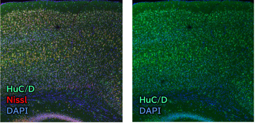
Species: Mouse
Site: Cerebral cortex
Sample: Frozen section
Antibody concentration: 1:1,500
Data by courtesy of
Dr. Miyata, Department of Applied Biology, Kyoto Institute of Technology
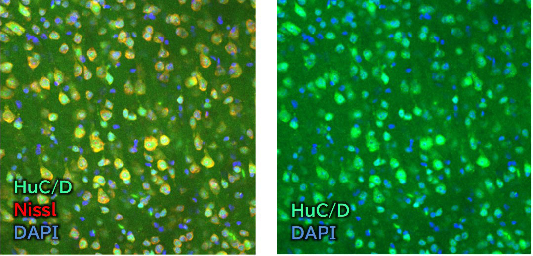
Species: Mouse
Site: Cerebral cortex (Layer V)
Sample: Frozen section
Antibody concentration: 1:1,500
Data by courtesy of
Dr. Miyata, Department of Applied Biology, Kyoto Institute of Technology
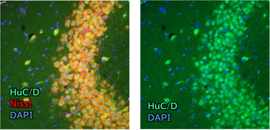
Species: Mouse
Site: Hippocampus (CA3)
Sample: Frozen section
Antibody concentration: 1:1,500
Data by courtesy of
Dr. Miyata, Department of Applied Biology, Kyoto Institute of Technology
References
- Szabo, A. et al.: Cell, 67(2), 325(1991).
HuD, a paraneoplastic encephalomyelitis antigen, contains RNA-binding domains and is homologous to Elav and Sex-lethal - Akamatsu, W. et al.: Proc. Nat. Acad. Sci. USA, 102(12), 4625(2005).
The RNA-binding protein HuD regulates neuronal cell identity and maturation
Product List
- Open All
- Close All
Anti HuC/D, Guinea Pig (Polyclonal)
For research use or further manufacturing use only. Not for use in diagnostic procedures.
Product content may differ from the actual image due to minor specification changes etc.
If the revision of product standards and packaging standards has been made, there is a case where the actual product specifications and images are different.
The prices are list prices in Japan.Please contact your local distributor for your retail price in your region.