Microglia Depletion Reagents
Microglia can be depleted using genetic or pharmacological methods, which are considered effective means for understanding the role of microglia. Although various methods have been developed, each has its own advantages and disadvantages, making it essential to choose the appropriate method based on an understanding of its characteristics. Fujifilm Wako supplies microglia depletion reagents using clodronate liposomes.
Methods of Microglia Depletion
Microglia are glial cells responsible for the immune functions of the central nervous system. They play a crucial role in phagocytosing foreign substances and dead cells, releasing chemokines and cytokines, monitoring neurons, and participating in synaptic pruning, thereby contributing significantly to the development and functions of the central nervous system. Additionally, in neurological and psychiatric disorders such as Alzheimer's disease and depression, microglia contribute to functional recovery and suppression of progression, but they can also cause brain inflammation, leading to disease onset and exacerbation.
Microglia depletion is an effective approach to elucidate their roles. Research is also being conducted on replacing microglia after depletion, as a potential therapeutic strategy for treating disorders. Several methods for depleting microglia, broadly categorized into genetic and pharmacological approaches, have been reported to date1). This article introduces methods for microglia depletion and the characteristics of each approach.
Genetic Deletion Models
Mutant genes necessary for microglia development (such as PU.1, TGF-β, and CSF-1R) have been utilized as models of microglial deficiency. More recently, a method has been employed to selectively deplete microglia by administering diphtheria toxin to transgenic mice that express the diphtheria toxin receptor specifically in microglia. Diphtheria toxin can cross the blood-brain barrier (BBB), conferring the advantage of enabling microglia depletion via intraperitoneal injection. However, a limitation of this method is that the depletion of microglia is not maintained for a prolonged period.
Pharmacological Treatment
Pharmacological methods for depleting microglia are easier to implement compared to genetic methods because they do not require the creation of transgenic models. The following three types of compounds are the main ones used to deplete microglia.
Clodronate Liposomes
Liposomes encapsulating clodronate are phagocytosed by microglia and macrophages. Once released into the cytoplasm, clodronate inhibits the mitochondrial ADP/ATP carrier, inducing cell death and thereby depleting microglia. While this method allows for rapid microglia depletion, it requires an intracerebral injection because liposomes cannot cross the BBB. Additionally, attention must be paid to potential side effects, such as the induction of inflammatory cytokines.
CSF-1R Inhibitors
CSF-1R is the receptor for colony-stimulating factor 1 (CSF-1) and is essential for the survival, proliferation, and differentiation of microglia and macrophages. Inhibiting CSF-1R can deplete microglia, with inhibitors such as PLX5622 and PLX3397 commonly used for this purpose. These inhibitors are notable for their ability to deplete microglia via oral administration and their high depletion efficiency. However, a drawback is the high cost of PLX5622 and PLX3397, which can increase the overall expense of experiments. Additionally, when administered orally, these drugs may also affect peripheral tissues, which requires careful consideration.
Ganciclovir
Ganciclovir (GCV) is structurally similar to nucleotides and functions as an antiviral agent by competitively inhibiting the uptake of substrates by viral DNA polymerase/reverse transcriptase. Intracerebroventricular injection of ganciclovir has been shown to deplete microglia, leading to its use as a microglia depletion agent. However, this method has several drawbacks: the requirement for intracerebroventricular injection due to ganciclovir’s inability to cross the BBB, the short duration of microglia depletion, and the induction of astrocyte activation. Despite the drawbacks, ganciclovir does not affect brain-associated macrophages (BAMs) or circulating bone marrow cells, making it an effective method for depleting microglia while minimizing effects on these cell types.
Microglia Remover
Microglia Remover is anionic liposomes suspension containing clodronic acid. When this product is phagocytosed by microglia as foreign substances, clodronic acid is released within the cells, causing cell death. By encapsulating clodronic acid in liposomes, it is taken up into microglia more efficiently than clodronic acid alone. Microglia remover (for Control) (sold separately, Product Number 131-19511) is also available as a control for experiments.
Features
- Microglia can be removed in approximately 1-3 days after administration
- Less expensive than commercial microglia removal feeds
Application Data
Microglia removal in mouse cerebral cortex
After inserting a 33 G needle into the mouse cerebral cortex, 1 µL of Microglia Remover (for Control) or Microglia Remover (this product) was injected, respectively, and sampled after 3 days.
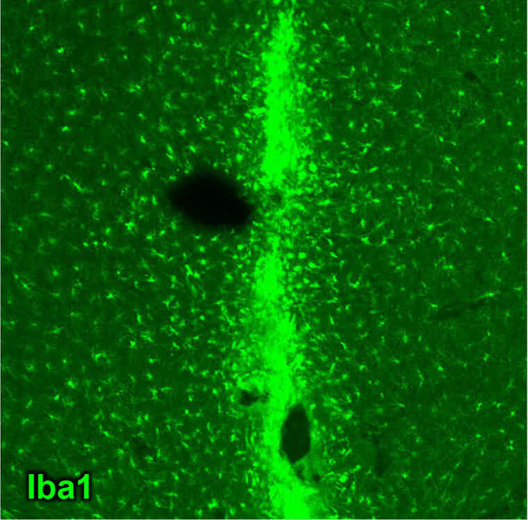
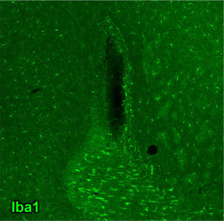
- Animal
- Mouse (C57BL6/J)
- Site
- Cerebral cortex
- Sample
- Frozen section
Antibody Concentration
- Primary Antibody
- Anti Iba1, Rabbit (for immunocytochemistry)
(Product Number 019-19741) 1:500 - Secondary Antibody
- Alexa Fluor® 488 AffiniPure Goat Anti-Rabbit IgG (H+L)
(Jackson ImmunoResearch, Product Number 111-545-144) 1:500
Dr. Miyata, Department of Applied Biology, Kyoto Institute of Technology
- [Result]
- In the control group, microglia (or macrophages) accumulated in large numbers at the site of injury.
On the other hand, the “Microglia Remover” group showed inhibition of the accumulation of microglia in the injured area and observed a decrease or regression in the number of microglia in the surrounding area.
References
- 1) Tahmasebi, F. and Barati, S.: Cell. Mol. Neurobiol., 43(6), 2459(2023).
The Role of Microglial Depletion Approaches in Pathological Condition of CNS
Product List
- Open All
- Close All
For research use or further manufacturing use only. Not for use in diagnostic procedures.
Product content may differ from the actual image due to minor specification changes etc.
If the revision of product standards and packaging standards has been made, there is a case where the actual product specifications and images are different.